
Image:Blotautoradiogram.png
|
|
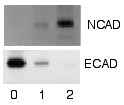
Example of a Western blot using radioactivity. This is an image of a sheet of film that was used to detect radioactively labeled Protein A on a sheet of nitrocellulose. Portions of two different gels (upper and lower panels) were transferred to the nitrocellulose. One was reacted with a primary antibody specific for the cell-to-cell adhesion protein N-cadherin, the other with anti-E-cadherin antibody. The three lanes (0, 1, 2) contained cell proteins from different times (days) after exposure to a chemical inducer of cell differentiation. The results show that when the cells differentiated they shifted from expressing one cadherin to the other.
Source: my personal image.
Uploaded for the Western blot page.
The copyright to this image is retained by John Schmidt (JWSchmidt).
Permission is granted to copy, distribute and/or modify this image under the terms of the Wikipedia GFDL, as indicated in the fine print at the bottom of this page.
Missing image Heckert_GNU_white.png | Permission is granted to copy, distribute and/or modify this document under the terms of the GNU Free Documentation License, Version 1.2 or any later version published by the Free Software Foundation; with no Invariant Sections, no Front-Cover Texts, and no Back-Cover Texts. Subject to disclaimers. |
If you do not want to use this image under the terms of the GFDL, you can alternatively use it under the terms of the following license:
| Missing image Stop_hand.png Image:Stop hand.png If this image was uploaded after May 19, 2005, it will soon be deleted without further warning.[1] (http://mail.wikipedia.org/pipermail/wikien-l/2005-May/023760.html) |
| Older images with this template will be considered for deletion. |
| This image is licensed under the Creative Commons Attribution NonCommercial ShareAlike 1.0 License:
http://creativecommons.org/licenses/by-nc-sa/1.0/ For the purposes of Wikipedia, this is a non-free license. |
File links
There are no pages that link to this file.